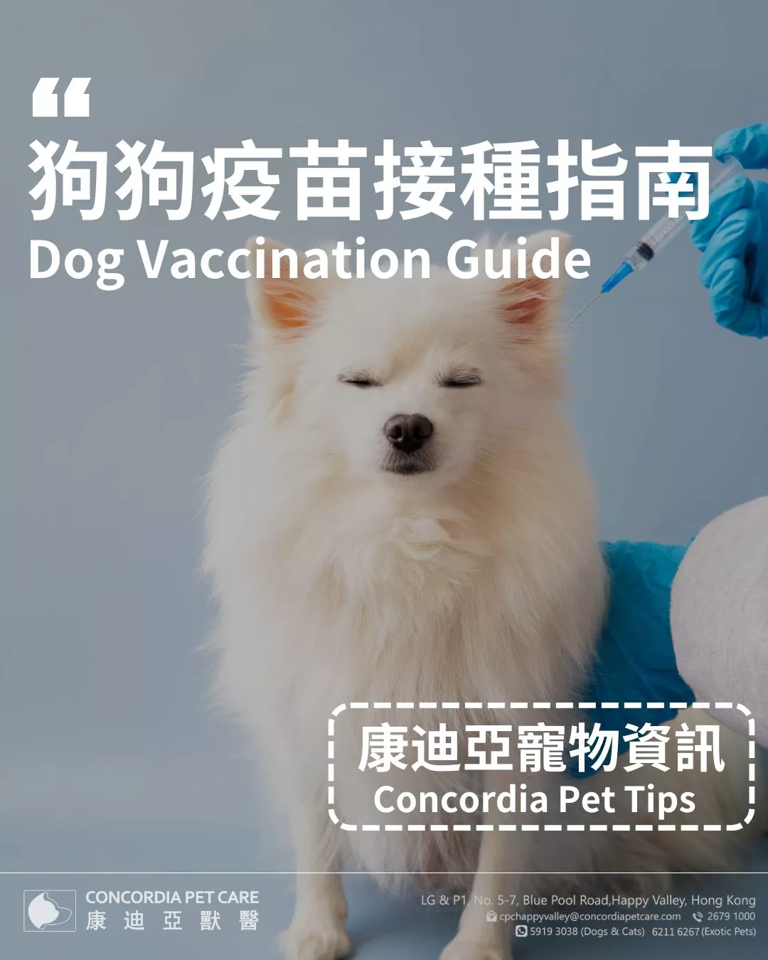
CONCORDIA NEWS
We will protect your pet with 100% care and professionalism!
 Concordia Pet Care
 2025-08-22


A diagnosis of mast cell tumor in dogs can be frightening, but early detection and the right plan make a real difference. In this guide, we’ll explain how to spot concerns, confirm a diagnosis, and choose care for a canine mast cell tumor.
A mast cell tumor dog case involves an overgrowth of mast cells, which are immune cells that normally help with allergy responses. When genetic changes drive uncontrolled growth, they form skin or subcutaneous masses that may release histamine and other chemicals, causing swelling, redness, and sometimes stomach irritation. Most arise on the skin, yet they can occur anywhere, including the spleen or liver in advanced situations.
Mast Cell Tumor vs. Mast Tumor in Dogs
“Mast cell tumor” is the correct medical term; “mast tumor” is a shorthand pet owners sometimes use. Both point to the same disease of mast cells. Your vet will use “mast cell tumor” on records, cytology, and pathology reports for clarity.
Some dog breeds show a higher risk for mast cell tumor. Reported predispositions include:
● Boxers, Pugs, and Boston Terriers
● Bulldogs, Labrador Retrievers, and Golden Retrievers
● Shar-Pei (tends to present aggressively)
Age is often middle to senior, but any dog can be affected, so new or changing skin lumps merit a check.
How Rare Is Mast Cell Tumor in Puppies?
A mast cell tumor diagnosis in a puppy is uncommon but possible. When it occurs in young dogs, behavior may be less aggressive. Still, prompt sampling and a tailored plan are essential to avoid progression. Early removal and staging help guide follow-up for a mast cell tumor detected in a puppy during routine exams.
Look for a solitary or multiple lump/s that wax and wane in size. This is common in dogs with mast tumor lesions that release histamine. Lumps can be firm or soft, hairless or haired, and may become red or puffy after handling.
Degranulation from mast cell tumor lesions in dogs can trigger vomiting, reduced appetite, diarrhea or black stools (suggesting GI ulceration), or lethargy. Sudden hives or facial swelling can also occur.
Ulceration on the lump, rapid regrowth after a previous removal, or enlarged nearby lymph nodes are concerning changes. Any mass that persists longer than two weeks or changes quickly, especially in breeds listed above, should be sampled.
Diagnosing a Suspected Mast Cell Tumor in Dogs
The quickest, least invasive first step is an FNA, where a veterinarian inserts a fine needle into the lump to collect cells for microscopic review. Under the microscope, canine mast cell tumor cells are typically round with distinct purple granules in their cytoplasm. This test can often confirm the diagnosis without immediate surgery, enabling the vet to plan the most appropriate next steps.
If surgery is planned or cytology is inconclusive, a tissue biopsy provides more detail. This allows a pathologist to assign a tumor grade, which predicts how aggressive the mast cell tumor in dogs may be and whether additional treatments beyond surgery are needed.
To determine if the disease has spread, your vet may check regional lymph nodes for abnormal cells, perform an abdominal ultrasound to look for lesions in the liver or spleen, and in some cases, use thoracic imaging to examine the chest cavity. This information helps shape a precise treatment plan and gives a clearer idea of the likely outcome.
Pathologists assign a grade to a canine mast cell tumor, which reflects how aggressive the tumor cells look and how likely they are to grow or spread. Understanding the grade helps vets recommend the right combination of surgery, follow-up, and other therapies.
● Low-grade: These tumors grow slowly, are less likely to spread, and often have excellent outcomes with complete surgical removal. Many patients live for years without recurrence.
● High-grade: These are more aggressive, with a higher risk of returning or metastasizing. Median survival is usually shorter, and multimodal therapy is often advised to manage the disease.
With timely FNAs, appropriate staging, and a tailored plan, outcomes for a mast cell tumor in canines are often far better than owners expect. Many dogs go on to live full, happy lives after treatment for mast cell tumor.
Surgery is the primary treatment when feasible. Vets typically aim for wide lateral margins and one fascial plane deep to reduce regrowth risk; tumor location (e.g., limbs, muzzle) can limit margins and influence recurrence. Costs reflect complexity (pre-op imaging, anesthesia time, reconstructive closure) and pathology fees.
Post-operative radiation is recommended when margins are incomplete or when tumors are unresectable due to anatomy. It is localized, delivered in planned fractions, and is particularly helpful for controlling residual microscopic disease around the surgical site.
For non-resectable, recurrent, or high-grade disease, systemic therapy can slow progression and improve comfort. Protocols may combine traditional chemotherapy with targeted options chosen by a veterinary oncologist, based on stage, grade, and patient tolerance.
When cure isn’t possible, the focus shifts to controlling itch, swelling, and GI irritation, along with pain relief and stomach-protective measures. This approach aims to keep patients comfortable at home and can meaningfully extend good-quality time.
Run your hands over your dog monthly, note any new masses, take a photo, and measure them with a ruler. Bring any lump that persists for >2 weeks or changes rapidly for an FNA.
After treating a mast tumor in dogs, scheduled rechecks (often every 3–6 months for the first two years) catch recurrences early and keep GI side effects in check. At Concordia Pet Care, we can set a personalized recheck timetable and reminder plan based on your dog’s grade and stage.
Keep a log of appetite, stools (watch for black or tarry), energy, and any swelling or redness over previous surgery sites. Call promptly if you see vomiting, dark stools, or a fast-growing new mass, especially in breeds prone to canine mast cell tumors.